What is Corneal Dystrophy?
Corneal dystrophy refers to a group of genetic and often progressive eye diseases characterized by the accumulation of abnormal material in at least one of the five layers of the cornea.
Most corneal dystrophies progress slowly and in both eyes. The age at which one can expect symptoms varies on a case-by-case basis.
Diagnosis of Corneal Dystrophy
An optometrist can usually detect the presence of corneal dystrophy with a slit lamp microscope during a regular eye exam. During this simple test the doctor shines a bright, thin beam of light into your eye. With the help of a slit lamp, discoloration or deformation of the cornea become visible. The exam can also show the build-up of abnormal material in the cornea.
The eye care professional will look into your family history of eye diseases, as corneal dystrophies are generally inherited. In some cases, genetic testing can reveal whether you are affected.
Symptoms of Corneal Dystrophy
People with corneal dystrophy may not experience any symptoms, while in others, the disorder may lead to significant vision impairment. Vision can be affected by the irregular shape of the cornea, as light is not bent correctly onto the retina. In addition, the accumulation of foreign material within the cornea can cause it to become foggy. In many cases, patients experience recurrent corneal erosion. This occurs when the top cell layer (the epithelium) detaches from the layer beneath.
Common symptoms for most corneal dystrophies include:
- blurred vision
- increased sensitivity to light (photophobia)
- the feeling of having a foreign object in the eye
- itching, pain, or severe discomfort
- vision loss
How Can Corneal Irregularities Be Treated?
Traditional soft contact lenses cannot correct vision, as the lens merely takes on the irregular shape of the eye surface. Similarly, regular eyeglasses provide improvement only for moderate astigmatism; however, in high irregular astigmatism, eyeglasses are not able to correct the condition.
Corneal erosion may be treated with medication, such as antibiotics, lubricating eye drops, or ointments, to repair damage to corneal tissue. Laser treatment may be applied, or, in severe cases, a corneal transplant may be required.
Rigid Gas Permeable (RGP) lenses for Corneal Dystrophies
RGPs can correct a patient’s vision since the inner surface of these lenses can be adapted to the irregular cornea, while the outer surface maintains a smooth round shape, thus correcting refraction at each point of the cornea.
Scleral Lenses for Corneal Dystrophies
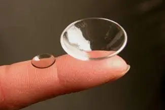
Scleral lenses offer clearer vision than RGP lenses as they vault over the corneal irregularity, forming a smooth dome-shaped surface. They are larger in diameter and rest on the sclera, the white part of the eye surrounding the cornea. The dome-shaped scleral lens spans over the cornea, creating an optically perfect surface with a liquid-filled vault underneath.
In addition, the lens is more comfortable to wear than most other lenses. The larger diameter prevents it from moving around on the eye, and the extra liquid between lens and cornea ensures that the eye remains well lubricated.
Types of Corneal Dystrophies
There are around 20 different types of corneal dystrophies, which can be divided into three distinct categories:
Anterior Corneal Dystrophies, which affect the outer layers of the cornea. These include Map-dot-fingerprint dystrophy, Lisch dystrophy, Meersman corneal dystrophy, Reis-Buckler corneal dystrophy and Thiel-Behnke corneal dystrophy.
Stromal Corneal Dystrophies, which affect the central or stromal layer of the cornea. These include granular corneal dystrophy, Lattice corneal dystrophy and macular corneal dystrophy.
Posterior Corneal Dystrophies, which affect the innermost layers of the cornea. These include congenital hereditary endothelial corneal dystrophy, Fuchs’ corneal dystrophy, posterior polymorphous corneal dystrophy and Schnyder crystalline corneal dystrophy.